T R Else, L Hacker, J Groehl, E V Bunce, R Tao, S E Bohndiek
Photoacoustic imaging (PAI) provides contrast based on the concentration of optical absorbers in tissue, enabling the assessment of functional physiological parameters such as blood oxygen saturation (sO2sO2). Recent evidence suggests that variation in melanin levels in the epidermis leads to measurement biases in optical technologies, which could potentially limit the application of these biomarkers in diverse populations
J Gröhl, T R Else, L Hacker, E V Bunce, P W Sweeney, S E Bohndiek
Accurate measurement of optical absorption coefficients from photoacoustic imaging (PAI) data would enable direct mapping of molecular concentrations, providing vital clinical insight. The ill-posed nature of the problem of absorption coefficient recovery has prohibited PAI from achieving this goal in living systems due to the domain gap between simulation and experiment. To bridge this gap, we introduce a collection of experimentally well-characterised imaging phantoms and their digital twins. This first-of-a-kind phantom data set enables supervised training of a U-Net on experimental data for pixel-wise estimation of absorption coefficients. We show that training on simulated data results in artefacts and biases in the estimates, reinforcing the existence of a domain gap between simulation and experiment. Training on experimentally acquired data, however, yielded more accurate and robust estimates of optical absorption coefficients. We compare the results to fluence correction with a Monte Carlo model from reference optical properties of the materials, which yields a quantification error of approximately 20%. Application of the trained U-Nets to a blood flow phantom demonstrated spectral biases when training on simulated data, while application to a mouse model highlighted the ability of both learning-based approaches to recover the depth-dependent loss of signal intensity. We demonstrate that training on experimental phantoms can restore the correlation of signal amplitudes measured in depth. While the absolute quantification error remains high and further improvements are needed, our results highlight the promise of deep learning to advance quantitative PAI.
L Hacker, H Wabnitz, A Pifferi, B W Pogue, J Pfefer and S E Bohndiek (2022) Nat Biomed Eng. 6 541-558.
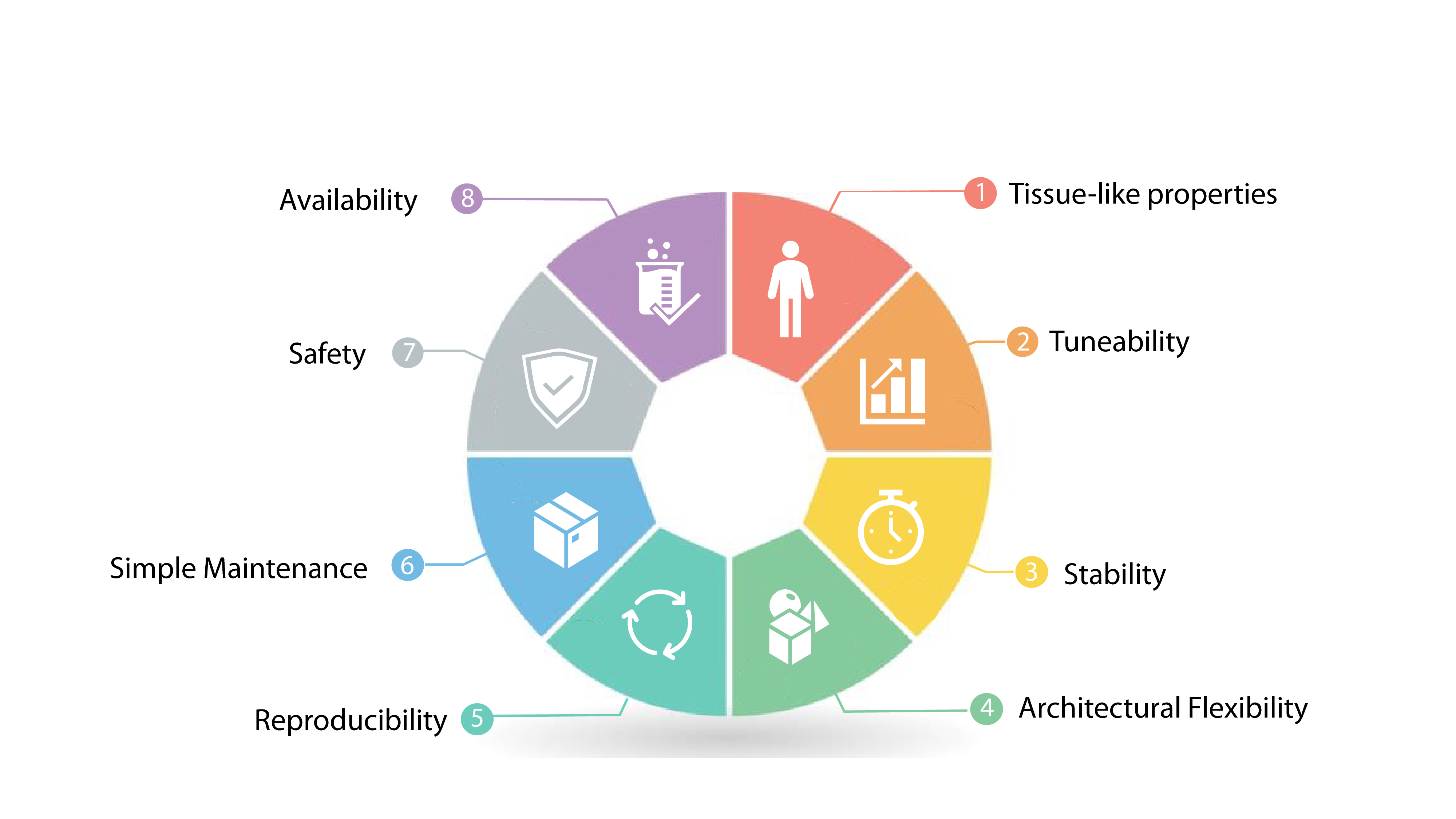
A lack of accepted standards and standardized phantoms suitable for the technical validation of biophotonic instrumentation hinders the reliability and reproducibility of its experimental outputs. In this Perspective, we discuss general criteria for the design of tissue-mimicking biophotonic phantoms, and use these criteria and state-of-the-art developments to critically review the literature on phantom materials and on the fabrication of phantoms. By focusing on representative examples of standardization in diffuse optical imaging and spectroscopy, fluorescence-guided surgery and photoacoustic imaging, we identify unmet needs in the development of phantoms and a set of criteria (leveraging characterization, collaboration, communication and commitment) for the standardization of biophotonic instrumentation.
L Hacker, J Joseph, A Ivory, M O Saed, B Zeqiri, S Rajagopal and S E Bohndiek (2021) IEEE Trans Med Imag. 40 (12) 3593-3603.
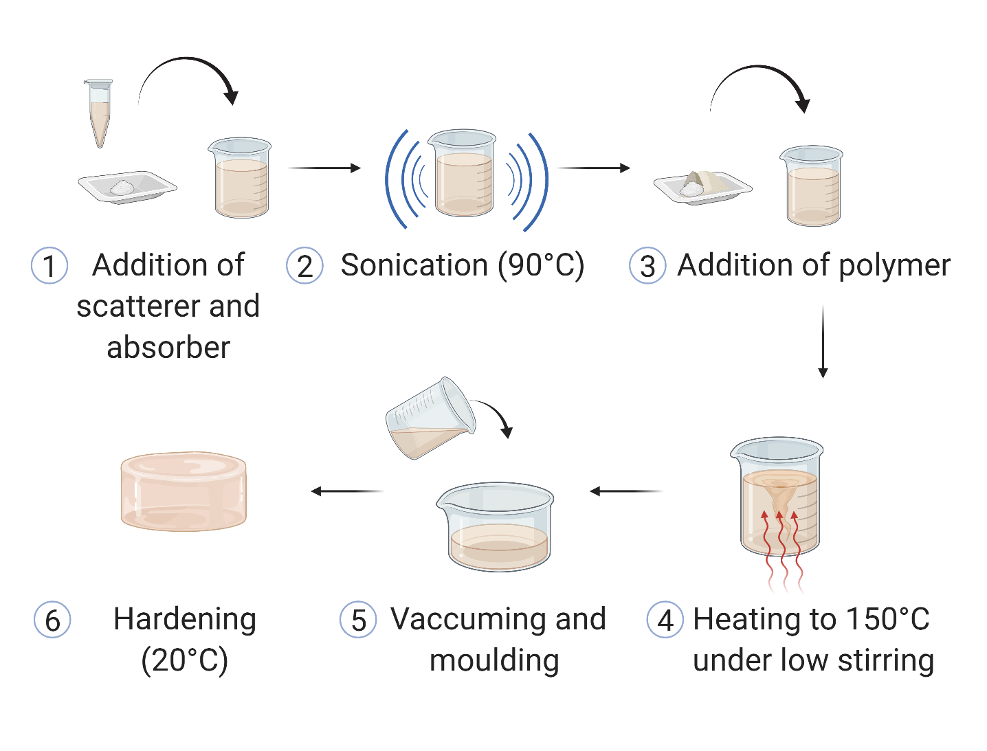
Photoacoustic imaging (PAI) standardisation demands a stable, highly reproducible physical phantom to enable routine quality control and robust performance evaluation. To address this need, we have optimised a low-cost copolymer-in-oil tissue-mimickingmaterial formulation. The base material consists of mineral oil, copolymer and stabiliser with defined Chemical Abstract Service numbers. Defined optical and acoustic properties were stable over almost a year and were minimally affected by recasting. The material showed high intra-laboratory reproducibility and good photo- and mechanical-stability in the relevant working range. The optimised copolymer-in-oil material represents an excellent candidate for widespread application in PAI phantoms, with properties suitable for broader use in biophotonics and ultrasound imaging standardisation efforts.
D J Waterhouse, W Januszewicz, S Ali, R C Fitzgerald, M di Pietro* and S E Bohndiek* (2021) Cancer Res 81 (12) 3415-3425.
The results of this pilot first-in-human clinical trial demonstrate the potential of spectral endoscopy to reveal disease-associated vascular changes and to provide high-contrast delineation of neoplasia in the esophagus.
C Williams, G S D Gordon, T D Wilkinson and S E Bohndiek (2019) ACS Photonics. 18;6(12):3132-3141. doi: 10.1021/acsphotonics.9b01196
Multispectral imaging is an exciting technology that can be applied to extract morphological (spatial) and biochemical (spectral) information from tumours. Typically, each multispectral imaging application requires a specific spectral range and number of spectral bands, which is challenging for conventional manufacturing processes. We present here a versatile, wafer-scale framework for producing highly efficient, narrowband and customisable transmissive multispectral imaging filter arrays. We envisage that the process can be used to fabricate custom multispectral imaging cameras, paving the way in the future for widespread application of the technology in biomedicine, including for endoscopic and intraoperative imaging.
J Yoon, J Joseph, D J Waterhouse, A S Luthman, G S D Gordon, M di Pietro, W Januszewicz, R C Fitzgerald and S E Bohndiek, Nature Communications, 10, Article number: 1902 (2019)
In this work, we demonstrate a hyperspectral endoscope (HySE) that simultaneously records intrinsically co-registered hyperspectral and standard-of-care white light images, which allows image distortions to be compensated computationally and an accurate hyperspectral data cube to be reconstructed as the endoscope moves in the lumen. Evaluation of HySE performance shows excellent spatial, spectral and temporal resolution and high colour fidelity. Application of HySE enables: quantification of blood oxygenation levels in tissue mimicking phantoms; differentiation of spectral profiles from normal and pathological ex vivo human tissues; and recording of hyperspectral data under freehand motion within an intact ex vivo pig oesophagus model. HySE therefore shows potential for enabling HSI in clinical endoscopy.
M. R. Tomaszewski, M Gehrung, J Joseph, I Quiros Gonzalez, J A Disselhorst and S E Bohndiek, Cancer Res. 78(20):5980-5991 (2018).
Oxygen Enhanced Optoacoustic Tomography (OE-OT) is a technique we developed and reported in 2017, exploiting gas challenge to provide high contrast insight into tumour vascular function. In this work, we show that OE-OT in combination with Dynamic Contrast Enhanced OT (DCE-OT), which relies on fluorescent contrast injection, can be used to accurately measure tumour oxygenation, maturity of the vascular network and the viability of the tumour tissue.
B Woodhams, L Ansel-Bollepalli, j Surmacki, H Knowles, L Maggini, M de Volder, M Atatüre and S E Bohndiek. (2018) Epub ahead of print at Nanoscale.
Nanodiamonds have demonstrated potential as powerful sensors in biomedicine. However, their translation into routine use requires a comprehensive understanding of their effect on the biological system being interrogated. In this paper we assessed the biological impact of graphitic and oxidized nanodiamond surfaces. We show for the first time that oxidized nanodiamonds possess improved biocompatibility compared to graphitic nanodiamonds in breast cancer cell lines, with graphitic nanodiamonds inducing higher levels of oxidative stress despite lower uptake.
Thore M. Bücking; Pim J. van den Berg; Stavroula Balabani; Wiendelt Steenbergen; Paul C. Beard; Joanna Brunker
In this work, researchers announce new advances in measuring blood flow velocity in deep tissue. Blood flow speed is a critical element in assessing tissue functionality as well as diagnosing diseases, and photoacoustic flowmetry (PAF) is already acknowledged as a promising technique for deep tissue measurement of blood flow velocity. The new work demonstrates successful use of a handheld ultrasound probe common in clinical settings, paving the way to explore the feasibility of measuring flow in a physiologically realistic situation.
M R Tomaszewski, I Quiros-Gonzalez, J O'Connor, O Abeyakoon, G Parker, K Williams, F Gilbert and S E Bohndiek (2017) Theranostics. 7 (11) 2900-2915.
Optoacoustic tomography (OT) is an emerging clinical imaging modality that provides static images of endogenous haemoglobin concentration and oxygenation. Here, we demonstrate oxygen enhanced (OE)-OT, exploiting an oxygen gas challenge to visualise the spatiotemporal heterogeneity of tumour vascular function.
A S N Luthman, S Dumitru, I Quiros-Gonzalez, J Joseph and S E Bohndiek. (2017) J. Biophotonics. 10 (6-7) 840-853.
Spectrally resolved detector arrays are an exciting new technology that allows spectral information to be recorded by conventional CMOS cameras (akin to those found in your smartphone). Here, we show that an SRDA can in fact be used to detect fluorescence signals for biomedical imaging applications.
D J Waterhouse, J Joseph, A A Neves, M di Pietro, K M Brindle, R C Fitzgerald and S E Bohndiek (2016) J. Biomed. Opt. 21(8), 084001.
In this work, we created a bimodal endoscope to reveal the binding of a near infrared fluorescent dye sprayed onto oesophageal tissue for detection of early malignancy.
G S D Gordon, J Joseph, S E Bohndiek and T D Wilkinson (2015) J Lightwave Technol. 33 16 3419-3425.
The characterisation of modal propagation in fibre bundles is performed here to achieve lensless focusing of imaging data. This is a first step towards performing phase retrieval and lensless focusing during clinical endoscopy using a fibre bundle.
S E Bohndiek, Laura S Sasportas, Steve Machtaler, Jesse V Jokerst, Sharon Hori and Sanjiv S Gambhir (2015) J Nucl Med. Accepted.
We were able to show here for the first time that optoacoustic imaging is able to detect both the vessel regression and normalisation that is commonly observed with anti-angiogenic therapy. This opens the possibility of using optoacoustic imaging to schedule combination therapy with cytotoxic drugs.
S E Bohndiek, A Wagadarikar, C L Zavaleta, D Van De Sompel, E Garai, J V Jokerst, S Yazdanfar and S S Gambhir (2013) Proc Natl Acad Sci USA 110 (30) 12408-12413
Recently, we developed a dedicated instrument for high throughput Raman spectroscopy, accelerating preclinical studies with this technique and hence improving the long-term prospects for clinical translation.
S E Bohndiek, M I Kettunen, D Hu, B W C Kennedy, J Boren, F A Gallagher and K M Brindle (2011) J Am Chem Soc A 133 (30) 11795-11801.
In this study, we demonstrated for the first time that vitamin C could be hyperpolarized and also used as a contrast agent to detect redox state noninvasively in living subjects.