T R Else, L Hacker, J Groehl, E V Bunce, R Tao, S E Bohndiek
Photoacoustic imaging (PAI) provides contrast based on the concentration of optical absorbers in tissue, enabling the assessment of functional physiological parameters such as blood oxygen saturation (sO2sO2). Recent evidence suggests that variation in melanin levels in the epidermis leads to measurement biases in optical technologies, which could potentially limit the application of these biomarkers in diverse populations
J Gröhl, T R Else, L Hacker, E V Bunce, P W Sweeney, S E Bohndiek
Accurate measurement of optical absorption coefficients from photoacoustic imaging (PAI) data would enable direct mapping of molecular concentrations, providing vital clinical insight. The ill-posed nature of the problem of absorption coefficient recovery has prohibited PAI from achieving this goal in living systems due to the domain gap between simulation and experiment. To bridge this gap, we introduce a collection of experimentally well-characterised imaging phantoms and their digital twins. This first-of-a-kind phantom data set enables supervised training of a U-Net on experimental data for pixel-wise estimation of absorption coefficients. We show that training on simulated data results in artefacts and biases in the estimates, reinforcing the existence of a domain gap between simulation and experiment. Training on experimentally acquired data, however, yielded more accurate and robust estimates of optical absorption coefficients. We compare the results to fluence correction with a Monte Carlo model from reference optical properties of the materials, which yields a quantification error of approximately 20%. Application of the trained U-Nets to a blood flow phantom demonstrated spectral biases when training on simulated data, while application to a mouse model highlighted the ability of both learning-based approaches to recover the depth-dependent loss of signal intensity. We demonstrate that training on experimental phantoms can restore the correlation of signal amplitudes measured in depth. While the absolute quantification error remains high and further improvements are needed, our results highlight the promise of deep learning to advance quantitative PAI.
L Hacker, H Wabnitz, A Pifferi, B W Pogue, J Pfefer and S E Bohndiek (2022) Nat Biomed Eng. 6 541-558.
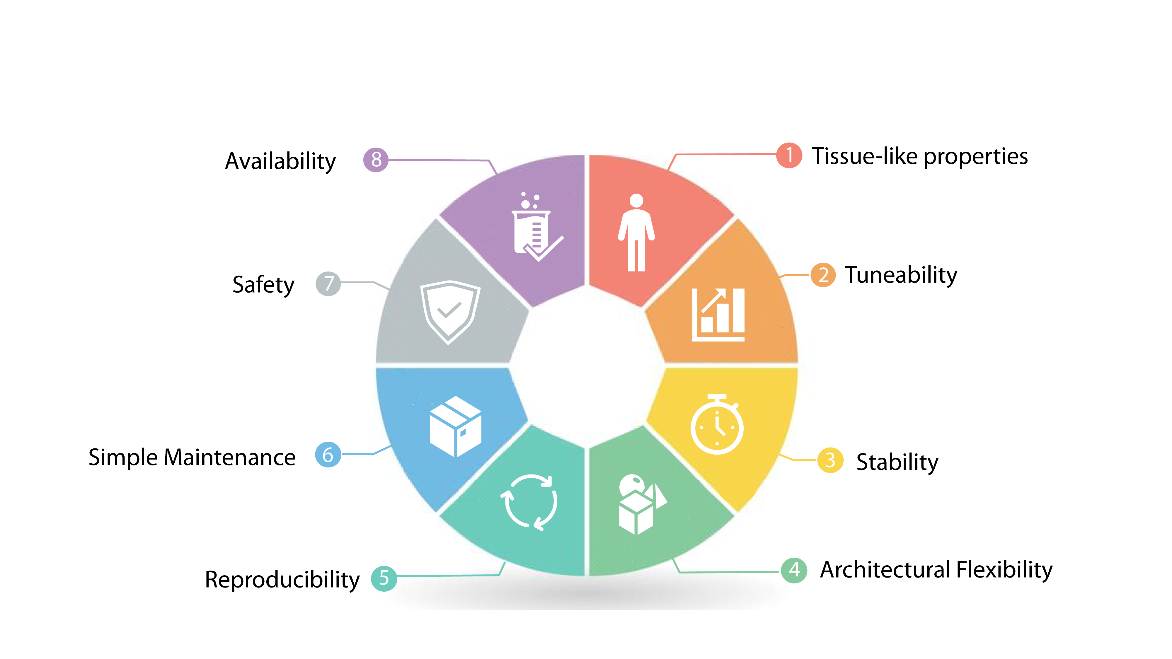
A lack of accepted standards and standardized phantoms suitable for the technical validation of biophotonic instrumentation hinders the reliability and reproducibility of its experimental outputs. In this Perspective, we discuss general criteria for the design of tissue-mimicking biophotonic phantoms, and use these criteria and state-of-the-art developments to critically review the literature on phantom materials and on the fabrication of phantoms. By focusing on representative examples of standardization in diffuse optical imaging and spectroscopy, fluorescence-guided surgery and photoacoustic imaging, we identify unmet needs in the development of phantoms and a set of criteria (leveraging characterization, collaboration, communication and commitment) for the standardization of biophotonic instrumentation.
L Hacker, J Joseph, A Ivory, M O Saed, B Zeqiri, S Rajagopal and S E Bohndiek (2021) IEEE Trans Med Imag. 40 (12) 3593-3603.
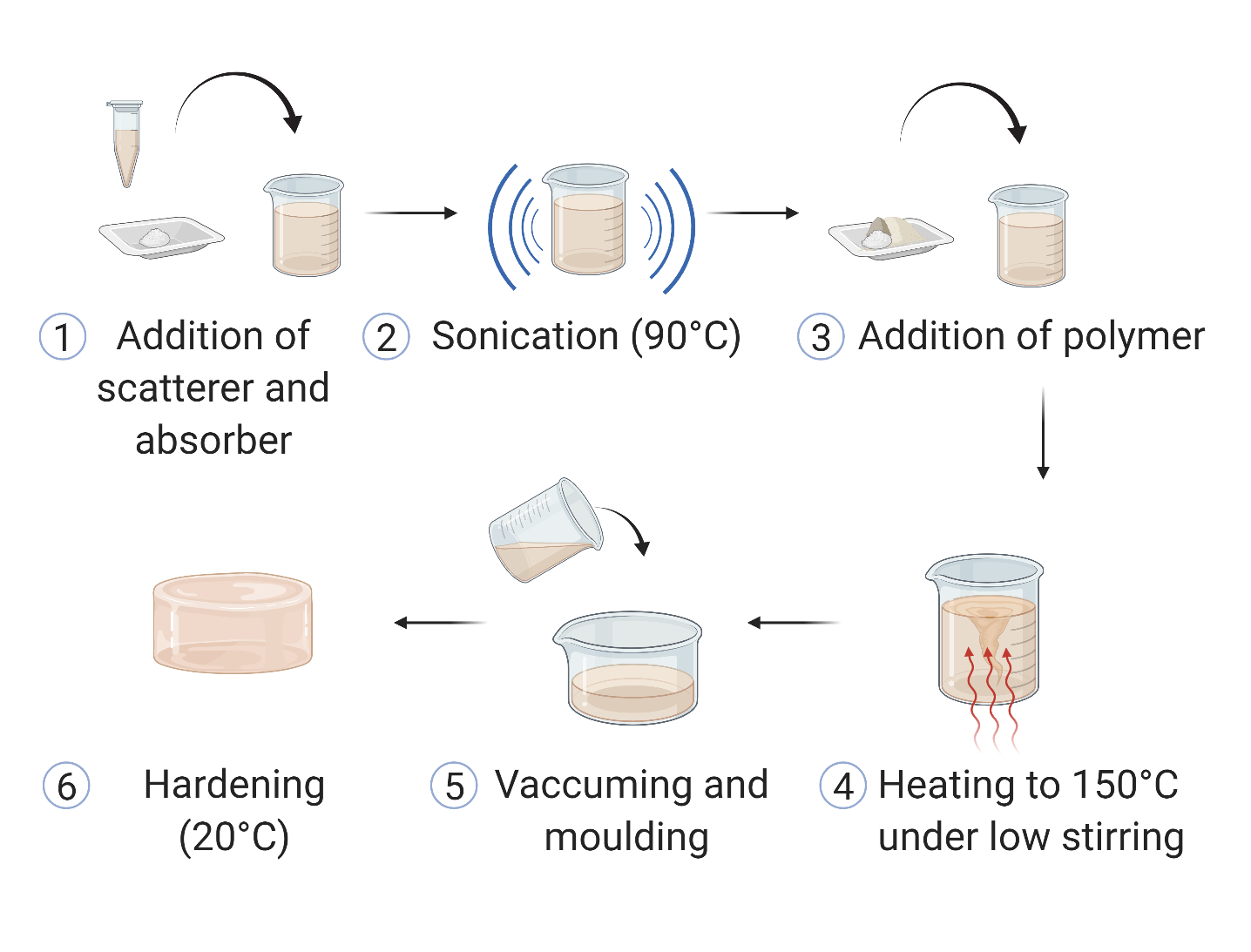
Photoacoustic imaging (PAI) standardisation demands a stable, highly reproducible physical phantom to enable routine quality control and robust performance evaluation. To address this need, we have optimised a low-cost copolymer-in-oil tissue-mimickingmaterial formulation. The base material consists of mineral oil, copolymer and stabiliser with defined Chemical Abstract Service numbers. Defined optical and acoustic properties were stable over almost a year and were minimally affected by recasting. The material showed high intra-laboratory reproducibility and good photo- and mechanical-stability in the relevant working range. The optimised copolymer-in-oil material represents an excellent candidate for widespread application in PAI phantoms, with properties suitable for broader use in biophotonics and ultrasound imaging standardisation efforts.
D J Waterhouse, W Januszewicz, S Ali, R C Fitzgerald, M di Pietro* and S E Bohndiek* (2021) Cancer Res 81 (12) 3415-3425.
The results of this pilot first-in-human clinical trial demonstrate the potential of spectral endoscopy to reveal disease-associated vascular changes and to provide high-contrast delineation of neoplasia in the esophagus.
S E Bohndiek, Laura S Sasportas, Steve Machtaler, Jesse V Jokerst, Sharon Hori and Sanjiv S Gambhir (2015) J Nucl Med. Accepted.
We were able to show here for the first time that optoacoustic imaging is able to detect both the vessel regression and normalisation that is commonly observed with anti-angiogenic therapy. This opens the possibility of using optoacoustic imaging to schedule combination therapy with cytotoxic drugs.